protocerebral bridge glomerulus 2-fan-shaped body layer 2-lateral accessory lobe-crepine neuron [FBbt_00111435]
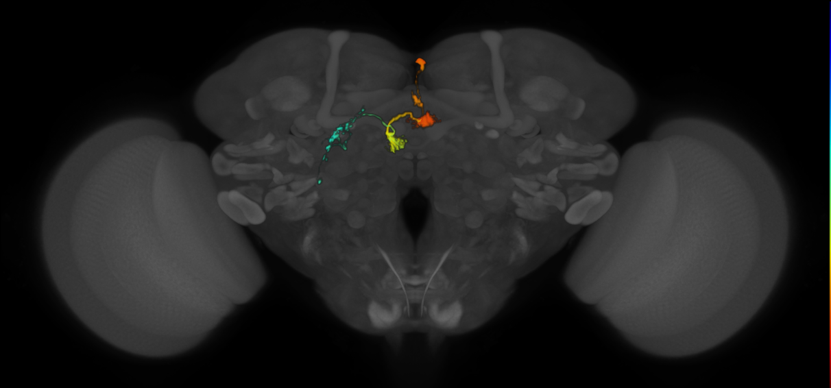
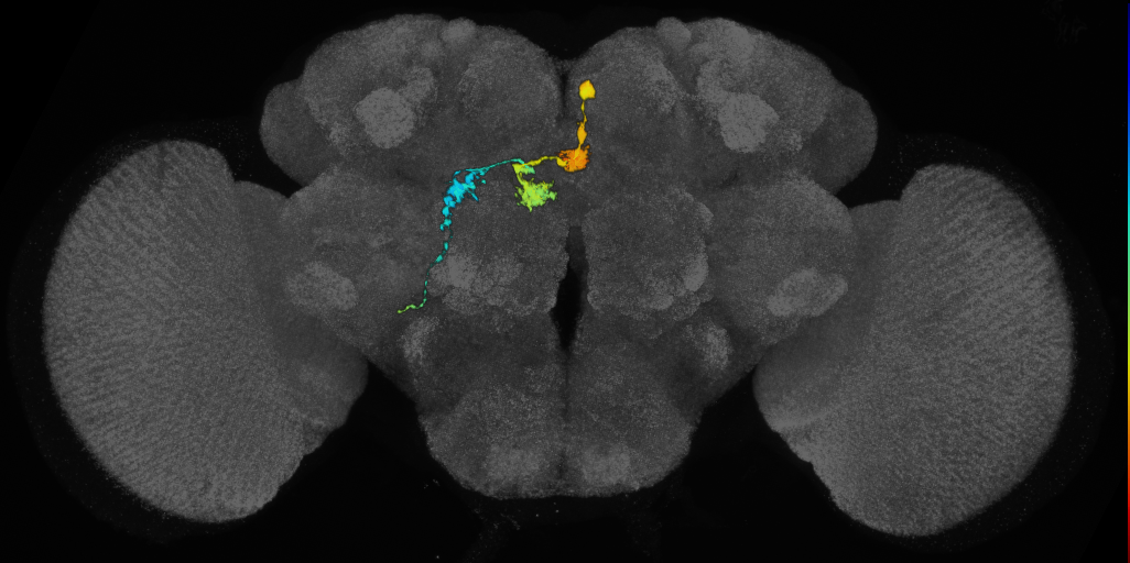
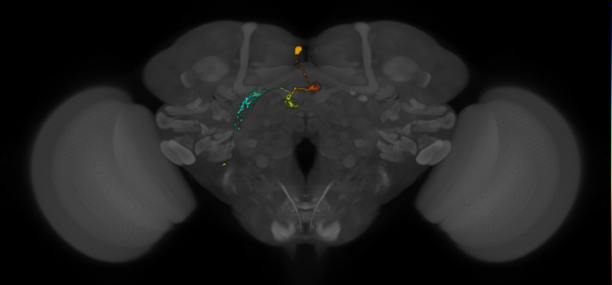
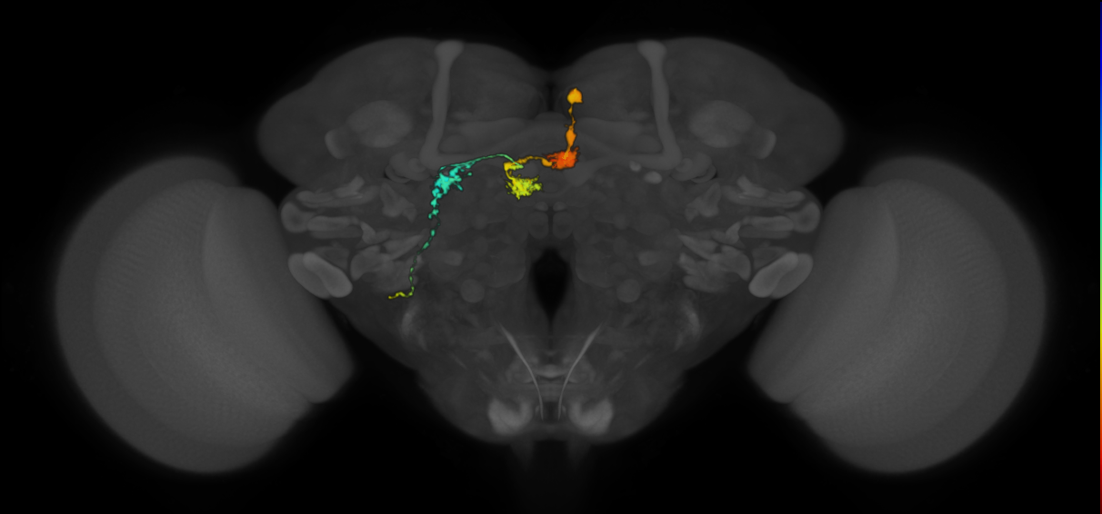
Adult protocerebral bridge 1 glomerulus-fan-shaped body layer 2-lateral accessory lobe-crepine (PFL1) neuron with dendritic arbors in protocerebral bridge (PB) glomerulus 2 and in the same contralateral fan-shaped body column targeted by the PFL1 cell of the contralateral PB glomerulus 6 (Lin et al., 2013; Hulse et al., 2020). Its axon terminals are in the contralateral lateral accessory lobe (Lin et al., 2013; Hulse et al., 2020). Due to the identification of an extra medial glomerulus by Wolff et al. (2020), the two ‘glomerulus 1’ cell types identified by Lin et al. (2013) are actually for the current glomerulus 1 and 2. This is likely to be the G2 type based on its more medial projection in the fan-shaped body compared to the G1 type, consistent with the G2 type in Hulse et al. (2020).
Note
This page displays the raw VFB json record for this term. Please use the link below to open the term inside the Virtual Fly Brain viewerExample Images
VFB Term Json
{
"term": {
"core": {
"iri": "http://purl.obolibrary.org/obo/FBbt_00111435",
"symbol": "",
"types": [
"Entity",
"Adult",
"Anatomy",
"Cell",
"Class",
"Nervous_system",
"Neuron"
],
"short_form": "FBbt_00111435",
"label": "protocerebral bridge glomerulus 2-fan-shaped body layer 2-lateral accessory lobe-crepine neuron"
},
"description": [
"Adult protocerebral bridge 1 glomerulus-fan-shaped body layer 2-lateral accessory lobe-crepine (PFL1) neuron with dendritic arbors in protocerebral bridge (PB) glomerulus 2 and in the same contralateral fan-shaped body column targeted by the PFL1 cell of the contralateral PB glomerulus 6 (Lin et al., 2013; Hulse et al., 2020). Its axon terminals are in the contralateral lateral accessory lobe (Lin et al., 2013; Hulse et al., 2020)."
],
"comment": [
"Due to the identification of an extra medial glomerulus by Wolff et al. (2020), the two 'glomerulus 1' cell types identified by Lin et al. (2013) are actually for the current glomerulus 1 and 2. This is likely to be the G2 type based on its more medial projection in the fan-shaped body compared to the G1 type, consistent with the G2 type in Hulse et al. (2020)."
]
},
"query": "Get JSON for Class",
"version": "44725ae",
"parents": [
{
"symbol": "PFL1",
"iri": "http://purl.obolibrary.org/obo/FBbt_00111433",
"types": [
"Entity",
"Adult",
"Anatomy",
"Cell",
"Class",
"Nervous_system",
"Neuron"
],
"short_form": "FBbt_00111433",
"label": "protocerebral bridge 1 glomerulus-fan-shaped body layer 2-lateral accessory lobe-crepine neuron"
}
],
"relationships": [
{
"relation": {
"iri": "http://purl.obolibrary.org/obo/RO_0013005",
"label": "receives_synaptic_input_throughout",
"type": "receives_synaptic_input_throughout"
},
"object": {
"symbol": "",
"iri": "http://purl.obolibrary.org/obo/FBbt_00003670",
"types": [
"Entity",
"Adult",
"Anatomy",
"Class",
"Nervous_system",
"Synaptic_neuropil",
"Synaptic_neuropil_subdomain"
],
"short_form": "FBbt_00003670",
"label": "protocerebral bridge glomerulus 2"
}
},
{
"relation": {
"iri": "http://purl.obolibrary.org/obo/RO_0000053",
"label": "bearer of",
"type": "bearer_of"
},
"object": {
"symbol": "",
"iri": "http://purl.obolibrary.org/obo/PATO_0000618",
"types": [
"Entity",
"Class"
],
"short_form": "PATO_0000618",
"label": "bilateral"
}
}
],
"xrefs": [],
"anatomy_channel_image": [
{
"anatomy": {
"symbol": "",
"iri": "http://virtualflybrain.org/reports/VFB_00005302",
"types": [
"Entity",
"Adult",
"Anatomy",
"Cell",
"Expression_pattern_fragment",
"Individual",
"NBLAST",
"Nervous_system",
"Neuron",
"VFB",
"has_image",
"NBLASTexp"
],
"short_form": "VFB_00005302",
"label": "Cha-F-300197"
},
"channel_image": {
"image": {
"template_channel": {
"symbol": "",
"iri": "http://virtualflybrain.org/reports/VFBc_00101567",
"types": [
"Entity",
"Individual",
"Template"
],
"short_form": "VFBc_00101567",
"label": "JRC2018Unisex_c"
},
"index": [],
"template_anatomy": {
"symbol": "",
"iri": "http://virtualflybrain.org/reports/VFB_00101567",
"types": [
"Entity",
"Adult",
"Anatomy",
"Individual",
"Nervous_system",
"Template",
"has_image"
],
"short_form": "VFB_00101567",
"label": "JRC2018Unisex"
},
"image_folder": "http://www.virtualflybrain.org/data/VFB/i/0000/5302/VFB_00101567/"
},
"channel": {
"symbol": "",
"iri": "http://virtualflybrain.org/reports/VFBc_00005302",
"types": [
"Entity",
"Individual",
"VFB"
],
"short_form": "VFBc_00005302",
"label": "Cha-F-300197-c"
},
"imaging_technique": {
"symbol": "Confocal",
"iri": "http://purl.obolibrary.org/obo/FBbi_00000251",
"types": [
"Entity",
"Class"
],
"short_form": "FBbi_00000251",
"label": "confocal microscopy"
}
}
},
{
"anatomy": {
"symbol": "",
"iri": "http://virtualflybrain.org/reports/VFB_00005302",
"types": [
"Entity",
"Adult",
"Anatomy",
"Cell",
"Expression_pattern_fragment",
"Individual",
"NBLAST",
"Nervous_system",
"Neuron",
"VFB",
"has_image",
"NBLASTexp"
],
"short_form": "VFB_00005302",
"label": "Cha-F-300197"
},
"channel_image": {
"image": {
"template_channel": {
"symbol": "",
"iri": "http://virtualflybrain.org/reports/VFBc_00017894",
"types": [
"Entity",
"Individual",
"Template",
"VFB"
],
"short_form": "VFBc_00017894",
"label": "JFRC2_template_c"
},
"index": [],
"template_anatomy": {
"symbol": "",
"iri": "http://virtualflybrain.org/reports/VFB_00017894",
"types": [
"Entity",
"Adult",
"Anatomy",
"Individual",
"Nervous_system",
"Template",
"VFB",
"has_image"
],
"short_form": "VFB_00017894",
"label": "adult brain template JFRC2"
},
"image_folder": "http://www.virtualflybrain.org/data/VFB/i/0000/5302/"
},
"channel": {
"symbol": "",
"iri": "http://virtualflybrain.org/reports/VFBc_00005302",
"types": [
"Entity",
"Individual",
"VFB"
],
"short_form": "VFBc_00005302",
"label": "Cha-F-300197-c"
},
"imaging_technique": {
"symbol": "Confocal",
"iri": "http://purl.obolibrary.org/obo/FBbi_00000251",
"types": [
"Entity",
"Class"
],
"short_form": "FBbi_00000251",
"label": "confocal microscopy"
}
}
},
{
"anatomy": {
"symbol": "",
"iri": "http://virtualflybrain.org/reports/VFB_00003932",
"types": [
"Entity",
"Adult",
"Anatomy",
"Cell",
"Expression_pattern_fragment",
"Individual",
"NBLAST",
"Nervous_system",
"Neuron",
"VFB",
"has_image",
"NBLASTexp"
],
"short_form": "VFB_00003932",
"label": "Cha-F-700198"
},
"channel_image": {
"image": {
"template_channel": {
"symbol": "",
"iri": "http://virtualflybrain.org/reports/VFBc_00017894",
"types": [
"Entity",
"Individual",
"Template",
"VFB"
],
"short_form": "VFBc_00017894",
"label": "JFRC2_template_c"
},
"index": [],
"template_anatomy": {
"symbol": "",
"iri": "http://virtualflybrain.org/reports/VFB_00017894",
"types": [
"Entity",
"Adult",
"Anatomy",
"Individual",
"Nervous_system",
"Template",
"VFB",
"has_image"
],
"short_form": "VFB_00017894",
"label": "adult brain template JFRC2"
},
"image_folder": "http://www.virtualflybrain.org/data/VFB/i/0000/3932/"
},
"channel": {
"symbol": "",
"iri": "http://virtualflybrain.org/reports/VFBc_00003932",
"types": [
"Entity",
"Individual",
"VFB"
],
"short_form": "VFBc_00003932",
"label": "Cha-F-700198-c"
},
"imaging_technique": {
"symbol": "Confocal",
"iri": "http://purl.obolibrary.org/obo/FBbi_00000251",
"types": [
"Entity",
"Class"
],
"short_form": "FBbi_00000251",
"label": "confocal microscopy"
}
}
},
{
"anatomy": {
"symbol": "",
"iri": "http://virtualflybrain.org/reports/VFB_00003932",
"types": [
"Entity",
"Adult",
"Anatomy",
"Cell",
"Expression_pattern_fragment",
"Individual",
"NBLAST",
"Nervous_system",
"Neuron",
"VFB",
"has_image",
"NBLASTexp"
],
"short_form": "VFB_00003932",
"label": "Cha-F-700198"
},
"channel_image": {
"image": {
"template_channel": {
"symbol": "",
"iri": "http://virtualflybrain.org/reports/VFBc_00101567",
"types": [
"Entity",
"Individual",
"Template"
],
"short_form": "VFBc_00101567",
"label": "JRC2018Unisex_c"
},
"index": [],
"template_anatomy": {
"symbol": "",
"iri": "http://virtualflybrain.org/reports/VFB_00101567",
"types": [
"Entity",
"Adult",
"Anatomy",
"Individual",
"Nervous_system",
"Template",
"has_image"
],
"short_form": "VFB_00101567",
"label": "JRC2018Unisex"
},
"image_folder": "http://www.virtualflybrain.org/data/VFB/i/0000/3932/VFB_00101567/"
},
"channel": {
"symbol": "",
"iri": "http://virtualflybrain.org/reports/VFBc_00003932",
"types": [
"Entity",
"Individual",
"VFB"
],
"short_form": "VFBc_00003932",
"label": "Cha-F-700198-c"
},
"imaging_technique": {
"symbol": "Confocal",
"iri": "http://purl.obolibrary.org/obo/FBbi_00000251",
"types": [
"Entity",
"Class"
],
"short_form": "FBbi_00000251",
"label": "confocal microscopy"
}
}
},
{
"anatomy": {
"symbol": "",
"iri": "http://virtualflybrain.org/reports/VFB_00011236",
"types": [
"Entity",
"Adult",
"Anatomy",
"Cell",
"Expression_pattern_fragment",
"Individual",
"NBLAST",
"Nervous_system",
"Neuron",
"VFB",
"has_image",
"NBLASTexp"
],
"short_form": "VFB_00011236",
"label": "Gad1-F-300066"
},
"channel_image": {
"image": {
"template_channel": {
"symbol": "",
"iri": "http://virtualflybrain.org/reports/VFBc_00101567",
"types": [
"Entity",
"Individual",
"Template"
],
"short_form": "VFBc_00101567",
"label": "JRC2018Unisex_c"
},
"index": [],
"template_anatomy": {
"symbol": "",
"iri": "http://virtualflybrain.org/reports/VFB_00101567",
"types": [
"Entity",
"Adult",
"Anatomy",
"Individual",
"Nervous_system",
"Template",
"has_image"
],
"short_form": "VFB_00101567",
"label": "JRC2018Unisex"
},
"image_folder": "http://www.virtualflybrain.org/data/VFB/i/0001/1236/VFB_00101567/"
},
"channel": {
"symbol": "",
"iri": "http://virtualflybrain.org/reports/VFBc_00011236",
"types": [
"Entity",
"Individual",
"VFB"
],
"short_form": "VFBc_00011236",
"label": "Gad1-F-300066-c"
},
"imaging_technique": {
"symbol": "Confocal",
"iri": "http://purl.obolibrary.org/obo/FBbi_00000251",
"types": [
"Entity",
"Class"
],
"short_form": "FBbi_00000251",
"label": "confocal microscopy"
}
}
}
],
"pub_syn": [
{
"synonym": {
"scope": "has_narrow_synonym",
"label": "PBL1-FBR2e,R3e>IDFPR-HB-lateral",
"type": ""
},
"pub": {
"core": {
"symbol": "",
"iri": "http://flybase.org/reports/FBrf0221742",
"types": [
"Entity",
"Individual",
"pub"
],
"short_form": "FBrf0221742",
"label": "Lin et al., 2013, Cell Rep. 3(5): 1739--1753"
},
"FlyBase": "",
"PubMed": "23707064",
"DOI": "10.1016/j.celrep.2013.04.022"
}
},
{
"synonym": {
"scope": "has_narrow_synonym",
"label": "PBR1-FBL2e,L3e>IDFPL-HB-lateral",
"type": ""
},
"pub": {
"core": {
"symbol": "",
"iri": "http://flybase.org/reports/FBrf0221742",
"types": [
"Entity",
"Individual",
"pub"
],
"short_form": "FBrf0221742",
"label": "Lin et al., 2013, Cell Rep. 3(5): 1739--1753"
},
"FlyBase": "",
"PubMed": "23707064",
"DOI": "10.1016/j.celrep.2013.04.022"
}
},
{
"synonym": {
"scope": "has_narrow_synonym",
"label": "PB slice 1-FB layer 2-IDFP HB-lateral neuron",
"type": ""
},
"pub": {
"core": {
"symbol": "",
"iri": "http://flybase.org/reports/Unattributed",
"types": [
"Entity",
"Individual",
"pub"
],
"short_form": "Unattributed",
"label": ""
},
"FlyBase": "",
"PubMed": "",
"DOI": ""
}
}
],
"def_pubs": [
{
"core": {
"symbol": "",
"iri": "http://flybase.org/reports/FBrf0227801",
"types": [
"Entity",
"Individual",
"pub"
],
"short_form": "FBrf0227801",
"label": "Wolff et al., 2015, J. Comp. Neurol. 523(7): 997--1037"
},
"FlyBase": "",
"PubMed": "25380328",
"DOI": "10.1002/cne.23705"
}
]
}
Feedback
Was this page helpful?
Glad to hear it! Please tell us how we can improve.
Sorry to hear that. Please tell us how we can improve.