ventrolateral domain of larval central nervous system [FBbt_00007705]
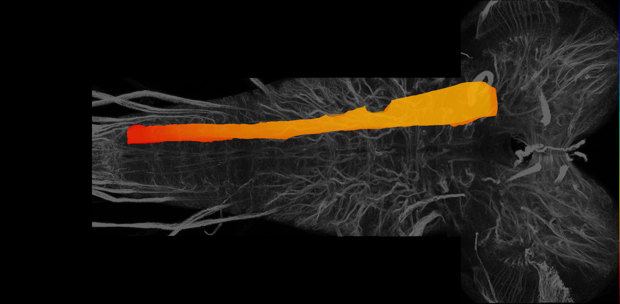
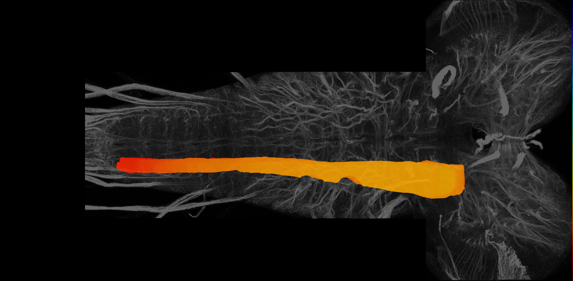
A ventro-laterally located longitudinal subdivision of the larval central nervous system spanning the gnathal, thoracic and abdominal neuromeres (Hartenstein et al., 2018). It is found at the same level as the ventromedial domain; ventral to the central domain (which contains the C1-3 fascicles) and the centrolateral domain (Hartenstein et al., 2018). Its medial boundary (with the ventromedial domain) is defined by the entry points of lineages 3,5,6 and 12 into the neuropil, which are at the same medio-lateral position as the C1-3 fascicles (Hartenstein et al., 2018). Anteriorly, it terminates posterior to the tritocerebrum, as the CITd and CITv (C1-3 fascicles) reach the lateral edge of the neuropil (Hartenstein et al., 2018). In thoracic neuromeres, this domain becomes enlarged during development and will give rise to the adult leg neuropils (Hartenstein et al., 2018).
Note
This page displays the raw VFB json record for this term. Please use the link below to open the term inside the Virtual Fly Brain viewerOpen ventrolateral domain of larval central nervous system in VFB
Example Images
VFB Term Json
{
    "term": {
        "core": {
            "iri": "http://purl.obolibrary.org/obo/FBbt_00007705",
            "symbol": "",
            "types": [
                "Entity",
                "Anatomy",
                "Class",
                "Larva",
                "Nervous_system"
            ],
            "short_form": "FBbt_00007705",
            "label": "ventrolateral domain of larval central nervous system"
        },
        "description": [
            "A ventro-laterally located longitudinal subdivision of the larval central nervous system spanning the gnathal, thoracic and abdominal neuromeres (Hartenstein et al., 2018). It is found at the same level as the ventromedial domain; ventral to the central domain (which contains the C1-3 fascicles) and the centrolateral domain (Hartenstein et al., 2018). Its medial boundary (with the ventromedial domain) is defined by the entry points of lineages 3,5,6 and 12 into the neuropil, which are at the same medio-lateral position as the C1-3 fascicles (Hartenstein et al., 2018). Anteriorly, it terminates posterior to the tritocerebrum, as the CITd and CITv (C1-3 fascicles) reach the lateral edge of the neuropil (Hartenstein et al., 2018). In thoracic neuromeres, this domain becomes enlarged during development and will give rise to the adult leg neuropils (Hartenstein et al., 2018)."
        ],
        "comment": []
    },
    "query": "Get JSON for Class",
    "version": "44725ae",
    "parents": [
        {
            "symbol": "",
            "iri": "http://purl.obolibrary.org/obo/FBbt_00007702",
            "types": [
                "Entity",
                "Anatomy",
                "Class",
                "Larva",
                "Nervous_system"
            ],
            "short_form": "FBbt_00007702",
            "label": "longitudinal domain of larval central nervous system"
        }
    ],
    "relationships": [],
    "xrefs": [],
    "anatomy_channel_image": [
        {
            "anatomy": {
                "symbol": "",
                "iri": "http://virtualflybrain.org/reports/VFB_00050351",
                "types": [
                    "Entity",
                    "Anatomy",
                    "Individual",
                    "Larva",
                    "Nervous_system",
                    "has_image"
                ],
                "short_form": "VFB_00050351",
                "label": "left ventrolateral subdivision of ventral nerve cord on L3 CNS template, Wood2018"
            },
            "channel_image": {
                "image": {
                    "template_channel": {
                        "symbol": "",
                        "iri": "http://virtualflybrain.org/reports/VFBc_00049000",
                        "types": [
                            "Entity",
                            "Individual",
                            "Template"
                        ],
                        "short_form": "VFBc_00049000",
                        "label": "L3 CNS template - Wood2018_c"
                    },
                    "index": [
                        95
                    ],
                    "template_anatomy": {
                        "symbol": "",
                        "iri": "http://virtualflybrain.org/reports/VFB_00049000",
                        "types": [
                            "Entity",
                            "Anatomy",
                            "Individual",
                            "Larva",
                            "Nervous_system",
                            "Template",
                            "has_image"
                        ],
                        "short_form": "VFB_00049000",
                        "label": "L3 CNS template - Wood2018"
                    },
                    "image_folder": "http://www.virtualflybrain.org/data/VFB/i/0005/0351/"
                },
                "channel": {
                    "symbol": "",
                    "iri": "http://virtualflybrain.org/reports/VFBc_00050351",
                    "types": [
                        "Entity",
                        "Individual"
                    ],
                    "short_form": "VFBc_00050351",
                    "label": "left ventrolateral subdivision of ventral nerve cord on L3 CNS template, Wood2018_c"
                },
                "imaging_technique": {
                    "symbol": "",
                    "iri": "http://purl.obolibrary.org/obo/FBbi_00000224",
                    "types": [
                        "Entity",
                        "Class"
                    ],
                    "short_form": "FBbi_00000224",
                    "label": "computer graphic"
                }
            }
        },
        {
            "anatomy": {
                "symbol": "",
                "iri": "http://virtualflybrain.org/reports/VFB_00050295",
                "types": [
                    "Entity",
                    "Anatomy",
                    "Individual",
                    "Larva",
                    "Nervous_system",
                    "has_image"
                ],
                "short_form": "VFB_00050295",
                "label": "right ventrolateral subdivision of ventral nerve cord on L3 CNS template, Wood2018"
            },
            "channel_image": {
                "image": {
                    "template_channel": {
                        "symbol": "",
                        "iri": "http://virtualflybrain.org/reports/VFBc_00049000",
                        "types": [
                            "Entity",
                            "Individual",
                            "Template"
                        ],
                        "short_form": "VFBc_00049000",
                        "label": "L3 CNS template - Wood2018_c"
                    },
                    "index": [
                        39
                    ],
                    "template_anatomy": {
                        "symbol": "",
                        "iri": "http://virtualflybrain.org/reports/VFB_00049000",
                        "types": [
                            "Entity",
                            "Anatomy",
                            "Individual",
                            "Larva",
                            "Nervous_system",
                            "Template",
                            "has_image"
                        ],
                        "short_form": "VFB_00049000",
                        "label": "L3 CNS template - Wood2018"
                    },
                    "image_folder": "http://www.virtualflybrain.org/data/VFB/i/0005/0295/"
                },
                "channel": {
                    "symbol": "",
                    "iri": "http://virtualflybrain.org/reports/VFBc_00050295",
                    "types": [
                        "Entity",
                        "Individual"
                    ],
                    "short_form": "VFBc_00050295",
                    "label": "right ventrolateral subdivision of ventral nerve cord on L3 CNS template, Wood2018_c"
                },
                "imaging_technique": {
                    "symbol": "",
                    "iri": "http://purl.obolibrary.org/obo/FBbi_00000224",
                    "types": [
                        "Entity",
                        "Class"
                    ],
                    "short_form": "FBbi_00000224",
                    "label": "computer graphic"
                }
            }
        }
    ],
    "pub_syn": [
        {
            "synonym": {
                "scope": "has_exact_synonym",
                "label": "larval ventrolateral neuropil domain",
                "type": ""
            },
            "pub": {
                "core": {
                    "symbol": "",
                    "iri": "http://flybase.org/reports/FBrf0237252",
                    "types": [
                        "Entity",
                        "Individual",
                        "pub"
                    ],
                    "short_form": "FBrf0237252",
                    "label": "Hartenstein et al., 2018, J. Comp. Neurol. 526(1): 6--32"
                },
                "FlyBase": "",
                "PubMed": "28730682",
                "DOI": "10.1002/cne.24287"
            }
        },
        {
            "synonym": {
                "scope": "has_narrow_synonym",
                "label": "ventrolateral subdivision of ventral nerve cord",
                "type": ""
            },
            "pub": {
                "core": {
                    "symbol": "",
                    "iri": "http://flybase.org/reports/Unattributed",
                    "types": [
                        "Entity",
                        "Individual",
                        "pub"
                    ],
                    "short_form": "Unattributed",
                    "label": ""
                },
                "FlyBase": "",
                "PubMed": "",
                "DOI": ""
            }
        }
    ],
    "def_pubs": [
        {
            "core": {
                "symbol": "",
                "iri": "http://flybase.org/reports/FBrf0237252",
                "types": [
                    "Entity",
                    "Individual",
                    "pub"
                ],
                "short_form": "FBrf0237252",
                "label": "Hartenstein et al., 2018, J. Comp. Neurol. 526(1): 6--32"
            },
            "FlyBase": "",
            "PubMed": "28730682",
            "DOI": "10.1002/cne.24287"
        }
    ]
}
Feedback
Was this page helpful?
Glad to hear it! Please tell us how we can improve.
Sorry to hear that. Please tell us how we can improve.